Proteins taken from a spherical virus and combined with pieces of DNA can form tubular nanostructures, researchers report. The finding could offer clues to how such molecules self-assemble.
In nature, the well-studied cowpea chlorotic mottle virus turns the leaves of the cowpea plant yellow, but it doesn’t harm its host. The virus has a 20-sided, spherical outer shell composed of RNA and viral proteins. It owes this geometry to the way that these two components weakly bind, says Adam Zlotnick of the University of Oklahoma Health Science Center in Oklahoma City.
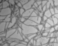
Zlotnick and his group combined the proteins with 500 base-pair-long pieces of double-stranded DNA in test tubes. Looking at the sample under transmission-electron microscopy, “we were delighted to see these beautiful tubes,” Zlotnick says.
The tubes are 17 nanometers in diameter and can be up to 5 micrometers long, depending on the ratio of DNA to viral protein. The negatively charged pieces of DNA—staggered in parallel along the length of the tube—act as an inner scaffold, attracting the positively charged proteins that form a tube’s wall.
The researchers describe the tubes in the March 1 Journal of the American Chemical Society.