50 years ago, X-rays provided an unprecedented look inside the brain
Excerpt from the September 1, 1973 issue of Science News
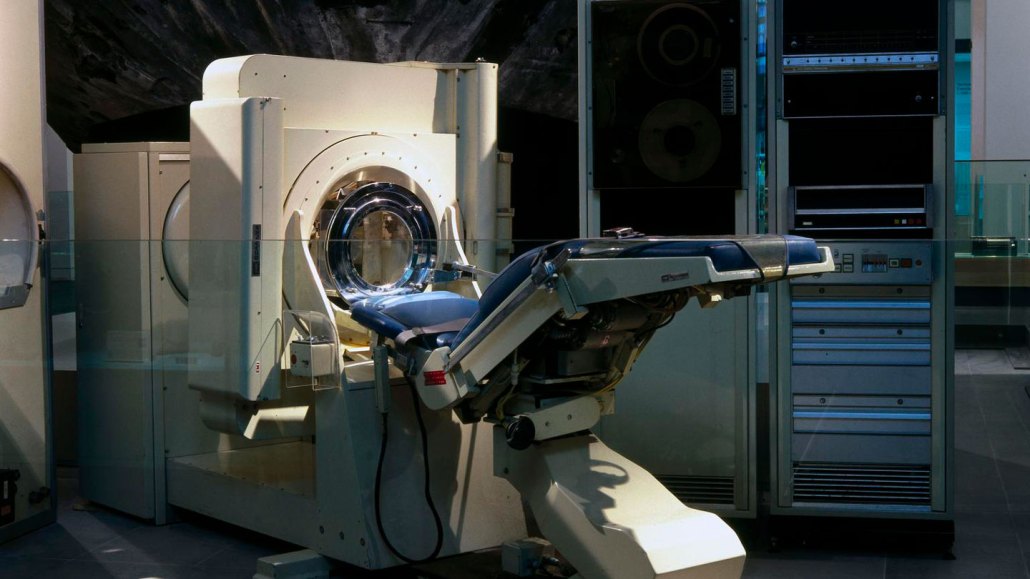
EMI brain scanners (one shown) developed in the 1970s paved the way for modern CT scanners, which can be used to examine the inside of the entire body.
Science Museum Group Collection © The Board of Trustees of the Science Museum (CC BY-NC-SA 4.0)

X-rays of the brain in thin cross sections — Science News, September 1, 1973
A new [device], already hailed by some physicians as the most important advance in X-ray diagnosis since its original development, promises to give a detailed new look at the inner brain.… The EMI device produces detailed information about a particular region of the brain with relatively little radiation exposure.
Update
That device was the first X-ray computed tomography, or CT, scanner. Today, the technology lets doctors and researchers peer inside not only the human brain, but also other organs, bones and even blood vessels. CT scanning has also become a useful tool in other areas of science, from archaeology to zoology (SN: 12/18/21 & 1/1/22, p. 44). For instance, the technology helped reveal why pumpkin toadlets are clumsy hoppers: Their inner ears may be too small to maintain good balance (SN: 7/16/22 & 7/30/22, p. 5). Sharper images made by a “photon-counting” CT scanner — approved last year by the U.S. Food and Drug Administration — could help resolve other mysteries too.