50 years ago, genes eluded electron microscopes

Excerpt from the September 2, 1972 issue of Science News

Yeast DNA transcription

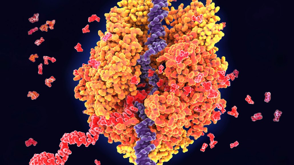
Scientists still can't directly see genes with electron microscopes, but combining the tool with the molecular scissors CRISPR/Cas9 has let researchers visualize genes being transcribed (illustrated) from DNA (blue) into RNA (red).

Juan Gaertner/Science Photo Library/Getty Images Plus

Cover of the September 2, 1972 issue of Science News

Visualizing Genes: The Possible Dream
– Science News, September 2, 1972

Molecular biologists can now visualize the larger structures of the cell, such as the nucleus and chromosomes, under the powerful electron microscope. But they have not been able to obtain images of genes (DNA) on the chromosomes. Nor have they been able to see RNA … or the intricate details of cell membranes, enzymes and viruses.

Update

Electron m­icroscopes have become much more powerful over the last 50 years. For instance, in 1981, biophysicist Jacques D­ubochet discovered that tiny biological structures super­cooled with ethane could be observed in their natural state under an electron microscope. That finding paved the way for cryo-electron micro­scopy, which scientists use to visualize proteins, viruses and bacteria at the molecular level (SN: 10/28/17, p. 6). Capturing detailed images of genes remains elusive, but scientists are inching closer. In 2021, researchers reported using an electron microscope and the molecular scissors CRISPR/Cas9 to visualize proteins transcribing DNA instructions for two genes into RNA.

Nikk Ogasa is a staff writer who focuses on the physical sciences for Science News. He has a master's degree in geology from McGill University, and a master's degree in science communication from the University of California, Santa Cruz.

More Stories from Science News on Tech