Microscopes have come a long way since 1665
Scopes deliver stunning cell images 350 years after Robert Hooke’s Micrographia
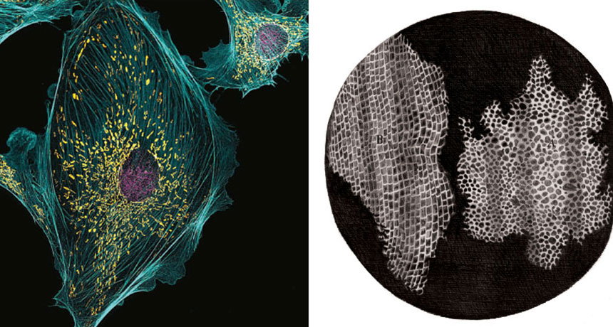
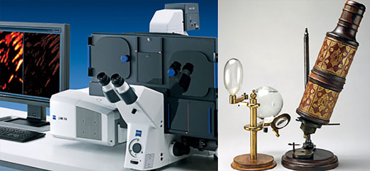
CENTURIES OF PROGRESS A Robert Hooke drawing from his 1665 book Micrographia (right) depicts little boxes in a slice of cork that he called cells. Today microscopes provide extraordinary views of cells, like these (left) from a cow.
Robert Markus; The Royal Society
In 1665, English scientist Robert Hooke published Micrographia, a book full of drawings depicting views through what was then a novel invention: the microscope. Peering at a slice of cork through a scope much like the one below (right), Hooke noticed small, boxlike partitions that he called cells (drawing above).
Now, 350 years later, cutting-edge microscopes enable biologists to study cells in extraordinary detail (SN: 6/15/13, p. 20). The above left photo, named an image of distinction in the 2015 Nikon Small World competition, shows cells that line the pulmonary artery of a cow; nuclei (purple), mitochondria (yellow) and structural fibers (blue) are clearly visible. Capturing the image required a super-resolution microscope (below, left) that Hooke could only have dreamed about in the 17th century.