New View: Speedy microscope takes fuller look at the nanoworld

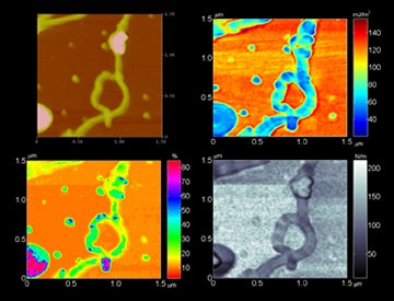
Although the atomic-force microscope is a workhorse for nanoscale measurements and manipulations, it’s neither the fastest nor the most informative of instruments. Used widely in biological and materials research, as well as in microelectronics manufacturing and other industries, the instrument provides minute topographical details of a sample but not much else.

NANOPORTRAITS. A single scan by an enhanced atomic-force microscope reveals (clockwise from upper left) the topography, adhesion, elasticity, and softness of a loop of carbon nanotubes Georgia Inst. Tech.

A team of engineers has now unveiled a radically revised version of the device. The inventors claim that it operates 100 times as fast as its conventional cousins do, raising the prospect that now-rare videos of molecular interactions could become routine. What’s more, as the new instrument examines the topography of an object, it can simultaneously measure other properties, such elasticity, stiffness, and stickiness, the scientists report.

“This is really powerful stuff,” claims mechanical engineer F. Levent Degertekin of the Georgia Institute of Technology in Atlanta. He and his colleagues describe their new device in the February Review of Scientific Instruments.

A standard atomic-force microscope, or AFM, probes a sample by means of a tiny cantilever with a sharp, downward-pointing tip (SN: 1/1/05, p. 12: Available to subscribers at Microscope goes mini). The instrument drags or taps the tip along a sample’s surface. By raising and lowering the cantilever to maintain a constant force on the tip, the instrument maps out the surface’s ups and downs.

“Right now, it’s a blind man’s world. We’re just feeling with the cantilever,” says physicist Thomas G. Thundat of Oak Ridge (Tenn.) National Laboratory, who works with AFM but was not on the design team for the new device.

Instead of a cantilever, the business end of the new-style AFM is a conventional tip mounted at the center of a circular membrane roughly the size of the period at the end of this sentence. The setup facilitates quick scanning because the membrane immediately flexes in response to an electrical signal within the device, Degertekin explains. Ordinary AFMs require more time to mechanically raise and lower the bulky apparatus that holds the cantilever.

Because the pliant membrane is also more responsive than the cantilever to forces imposed by a sample’s surface, its motions yield information about properties that affect those forces, Degertekin adds.

“If you’re going to look at nanostructures, you want a tool that will do everything,” says team member Calvin F. Quate of Stanford University, who coinvented the AFM in 1986. The membrane-based probe could be that all-purpose tool, he adds.

The next challenge is to run the new AFM in water to show whether it’s applicable to biological studies, comments AFM microscopist Paul K. Hansma of the University of California, Santa Barbara.

That shouldn’t be a problem, Degertekin says, because versions of the new AFM’s membrane have already served as ultrasound sensors in liquids.

More Stories from Science News on Tech