See these dazzling images of a growing mouse embryo

A new microscope lets scientists peek into the mysterious process of mammalian development

mouse embryo developing

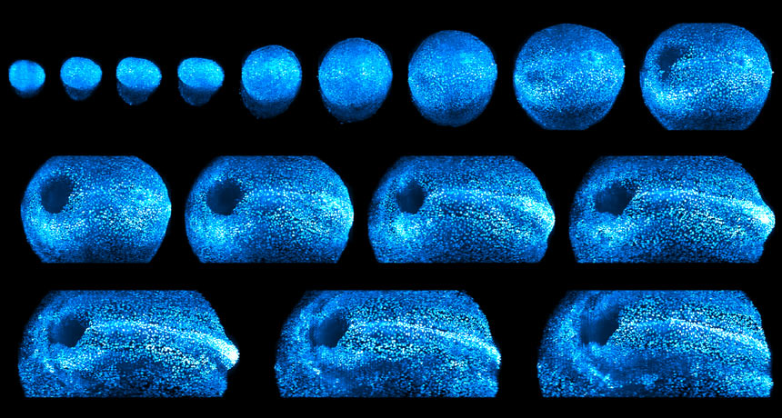
LIGHT THE WAY  A new microscope uses laser light to image growing mammal embryos. Scientists used the instrument to track a mouse embryo (seen in false color) as it developed over two days.

K. McDole et al/Cell 2018

A new microscope is giving researchers an unprecedented view of how mammals are built, cell by cell.

Light sheet microscopes use ultrathin laser beams to illuminate sections of a specimen while cameras record those lit-up sections. Previous iterations of the device have captured detailed portraits of living zebra fish and fruit fly embryos as they develop. Kate McDole, a developmental biologist at Howard Hughes Medical Institute’s Janelia Research Campus in Ashburn, Va., and colleagues used a new-and-improved version to monitor the development of a larger, more complex organism: the mouse.

Algorithms in the microscope tracked 6-day-old mouse embryos in real time over roughly two days, keeping the device focused on the cell clusters as they grew. A suite of computer programs used the data — about a million images per embryo — to map the life history of each embryo’s every cell, the team reports October 11 in Cell. The result: dazzling views of mouse organs taking shape.

As an embryo rapidly expands in size, the gut starts to form when part of the embryo collapses into a craterlike hole. And a structure that eventually forms the brain and spinal cord, called a neural tube, appears like a comet shooting across the night sky. Researchers also captured the first beats of heart cells.

GROWTH SPURT Scientists used a new laser-powered microscope to peek inside a growing mouse embryo (shown in false color in the first clip). Over two days, the team saw the formation of the mouse’s gut (dark blue hole that appears in the developing embryo) and neural tube (white line), which becomes the brain and spinal cord. Probing deeper into the embryo revealed newly developed heart cells’ first rhythmic beats (second clip).

“These are processes no one has been able to watch before,” McDole says. Seeing the gut form in minutes was stunning. “We never expected it to be that fast or that dramatic. It’s not like you can Google these things.”

Such imaging technology may help resolve a long-standing mystery: how mammals develop from single cells to fully formed multicellular embryos. That information is crucial for scientists attempting to grow human organs (SN: 3/6/04, p. 155) or fix defects that arise in embryos during pregnancy, McDole says.

“If you took all the building materials for a house and threw them in a pile, you don’t magically get a house. Contractors use plans to build the house,” she says. The same thing applies to human health, she says, so “being able to see how embryos actually make organs is a huge step forward.”

Cassie Martin is a deputy managing editor. She has a bachelor's degree in molecular genetics from Michigan State University and a master's degree in science journalism from Boston University.

More Stories from Science News on Life

From the Nature Index

Paid Content