New microscope techniques give deepest view yet of living cells

mouse embryonic fibroblast cell

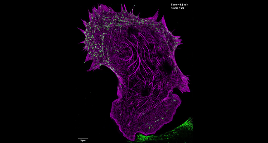
A new microscopy technique called nonlinear SIM gives a better look at how the cell’s internal scaffolding, or cytoskeleton, changes over time. Shown is a still image from a video of cytoskeletal proteins in a mouse embryonic fibroblast cell. 

D. Li et al/Science 2015

Two new microscopy techniques are helping scientists see smaller structures in living cells than ever glimpsed before.

Scientists can now view structures just 45 to 84 nanometers wide, Nobel prize-winning physicist Eric Betzig of the Howard Hughes Medical Research Institute’s Janelia research campus in Ashburn, Va., and colleagues report in the Aug. 28 Science. The techniques beat the previous resolution of 100 nanometers and shatters the 250 nanometer “diffraction barrier,” imposed by the bending of light.

Using other tricks to improve the super-resolution methods also allowed the researchers to take ultraquick pictures with less cell-damaging light than before. As a result, scientists can watch sub-second interactions within cells, revealing new insights into how cells work.

This video, taken with the nonlinear SIM techniques that can reveal structures as small as 84 nanometers, shows relationships between the actin cytoskeleton (red-orange) and pockets called endosomes (green) that cells use to internalize materials. D. Li et al/ Science 2015

Tina Hesman Saey is the senior staff writer and reports on molecular biology. She has a Ph.D. in molecular genetics from Washington University in St. Louis and a master’s degree in science journalism from Boston University.

More Stories from Science News on Life

From the Nature Index

Paid Content