Primates’ eyes view scenes like a seamless patchwork quilt, a study appearing online April 7 in PLoS Biology shows. This underappreciated visual coordination may contribute to why primates, including humans, can see with such precision, says study coauthor Jeffrey Gauthier.
More than a million nerve cells in each primate retina collect information in the form of light and transmit that information as signals to the brain. Each of these retinal ganglion cells views just a small corner of the world, called the receptive field, as if gazing out of a tiny airplane window.
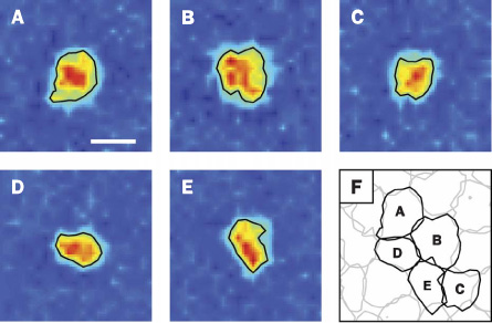
Earlier studies looking at one cell at a time found that each cell responded to light coming from an irregularly shaped blob of space. These jagged pieces could cover some places multiple times and others not at all, leading researchers to think that primates’ visual systems rely on averaging the input to make a complete view of the world. Such unevenness could lead to blind spots, where coverage is lacking, and to blurry vision in places of excess coverage, Gauthier, of the Salk Institute for Biological Studies in La Jolla, Calif., and his colleagues say in their paper.
In the new study, the team looked at hundreds of cells simultaneously to see how the retina’s cells work together. The researchers found that the irregularly shaped receptive fields of individual nerve cells nestled together perfectly, like a massive jigsaw puzzle.
The researchers identified the exact location and shape of the nerve cells’ receptive fields in two species of Macaca monkey retinas by flashing complex light patterns, which looked like television snow, to the retinal cells. The researchers used electrodes to tell when each individual nerve cell was activated.
“Everything the brain sees and learns from the retina, we can also see and learn,” Gauthier says.
Activation was then traced back to individual pixels of light from the snowy scene. By matching the pixels of light to each activated nerve cell, the researchers knew the size and shape of each cell’s receptive field.
To make sure the receptive fields were truly in lockstep, the researchers performed another test. When a computer program rotated each receptor field 180 degrees, gaps and overlaps appeared, presumably ruining the coordination between the receptor fields.
Researchers don’t yet know how the cells manage to form this intricate coordination. Neuroscientist Richard Masland of Harvard Medical School says, “A great remaining question is how this tiling is created developmentally.” Gauthier suggests that the cells may rely on visual input to shape the receptive fields.