Scientists spy on the secret inner life of bacteria
New images uncover mysterious structures inside microbes, including mini towers, fishhooks, train tracks and horseshoes

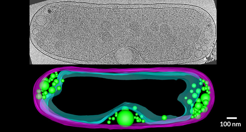
UNDER THE SKIN Researchers have discovered mysterious structures, such as these bubblelike compartments called vesicles that sit between the cell membrane (cyan) and the cell wall (purple) of the bacterium Halothiobacillus neapolitanus (cryotomogram, top; 3-D reconstruction, bottom).
M.J. Dobro/Journal of Bacteriology 2017
On the surface, bacteria may appear bland. But there’s more going on inside than meets the eye, new research is revealing.
For many years, scientists thought that bacteria didn’t have internal structures and were basically “bags of enzymes,” says structural and cell biologist Martin Warren of the University of Kent in England.
Now, one group of researchers has described a rich collection of mysterious structures and compartments within bacteria. No one knows the function of the constructs, the researchers report online June 12 in the Journal of Bacteriology, but they must be important for bacteria to spend so much energy building them.
A different team of scientists presents the first atomic-scale look at a complete bacterial microcompartment in the June 23 Science. Microcompartments are protein shells that bacteria use to keep certain chemical reactions separate from the rest of the cell. Knowing how the microcompartment is assembled could have important applications in biotechnology and medicine, the researchers say.
Inner sanctum
Bacteria use protein shells called microcompartments to concentrate important chemical reactions in one place. Now researchers have constructed a complete 3-D model of a microcompartment from a saltwater bacterium called Haliangium ochraceum. Hexagon-shaped structures composed of the BMC-H protein (blue) and BMC-T protein (green) combine with pentagons made of BMC-P protein (yellow) to create the shell. It is the largest protein structure, besides viruses, that scientists have ever viewed at atomic-level resolution.

The two studies demonstrate how complex bacteria really are, says Warren, who wasn’t involved in either. “They’re both fantastic pieces of work. If anybody ever thought microbiology was boring, they should have a look at this.”
Since the 1950s, biologists have known that photosynthetic cyanobacteria make microcompartments, called carboxysomes, which house an important photosynthesis enzyme. About 20 percent of bacteria — even ones that don’t do photosynthesis — have the genes for making similar microcompartments. Many of those tiny chambers appear only when bacteria encounter certain molecules they can use for food. For instance, some pathogenic bacteria form microcompartments to help them digest mucus from people’s intestines, Warren says.
Subscribe to Science News
Get great science journalism, from the most trusted source, delivered to your doorstep.
Many researchers have worked to engineer microcompartments to make drugs, industrial chemicals or biofuels. That’s been difficult because scientists didn’t fully understand the structures’ construction. They knew that proteins snapped together to make pentagon- and hexagon-shaped units, but didn’t know how the subunits came together in multi-sided spheres. “It’s like playing with Lego building blocks, but not understanding how the bricks fit together,” says Danielle Tullman-Ercek, a synthetic biologist at Northwestern University in Evanston, Ill.
In addition, the individual units are curved, with a cup on one side and a bulge on the other, says Michael Prentice, a medical microbiologist at University College Cork in Ireland. But it wasn’t clear whether the cup or the bulge went on the inside of the microcompartment.
Now, in the Science paper, researchers led by structural biologist Cheryl Kerfeld have used a combination of X-ray crystallography and cryo-electron microscopy to examine microcompartments from the bacterium Haliangium ochraceum and answer those questions. The cups cover the outsides of the microcompartments, like dimples on a golf ball, says Kerfeld of Michigan State University in East Lansing and the Lawrence Berkeley National Laboratory in California.
Story continues below image
Eiffel edifice
A freshwater bacterium called Prosthecobacter debontii studs its surface with appendages resembling mini Eiffel towers. A cryotomogram (left and upper left inset), and a 3-D model of the side view (right, gold) and top view (gold, inset) of the structure are shown. Scientists don’t know how the complex appendages are used.

Kerfeld’s team also found that the subunits’ sides can both contact each other edge-to-edge to make flat sheets and can join up at an angle, making a curved surface. “We couldn’t have predicted this,” Kerfeld says. Some of the hexagon-shaped subunits stack on top of each other giving the sphere an irregular appearance, the researchers found. The team is still exploring how the orientation of the subunits affects the compartment’s function.
Microcompartments aren’t the only mysterious structures inside bacteria. Over the last decade, structural cell biologist Grant Jensen of Caltech and colleagues have used electron cryotomography to make more than 15,000 images of 88 species of bacteria. The technique involves flash freezing cells so quickly that water molecules can’t form crystals. Cells are preserved as they are in life, allowing researchers to see what they really look like inside.
Jensen’s team found structures resembling the Eiffel Tower, fishhooks, horseshoes, railroad tracks, filaments and meshes. Many different compartments within the bacterial cells or between the cell membrane and the bacteria’s cell wall also became apparent.
“We were discovering them faster than we could identify them,” Jensen says. His team published the images in the Journal of Bacteriology because “we thought it would be best for the progress of science to show them to everybody.” He hopes crowdsourcing the images with other researchers will help solve the mysteries of what these structures are and what they do.


