Turn Your Head and Roar

Can diagnosing disease in fossils shed light on modern maladies?

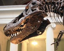
In one of the early scenes of Jurassic Park, the 1993 film in which dinosaurs were resurrected from their DNA, paleobiologist Ellie Sattler leaves her jeep during a guided tour to assist the park veterinarian, who is tending an obviously sick Triceratops.

All skulls from mature Tyrannosaurus rex, such as the Field Museum’s Sue, have holes in the rear-lower jaw that may be signs of long-term infections or bite marks from other members of the species. Rega

Photos (left) and X-ray images (right) show lesions in navicular bones from horses trapped in the La Brea tar pits (top row) and from modern horses (bottom row). Thompson

The Tyrannosaurus rex Sue’s many afflictions included a broken left rib that healed with a movable pseudojoint (top) and a long-term infection around a bone in the lower left leg (arrow, bottom). Thompson

The animal’s symptoms: Every 6 weeks it became disoriented, lost its sense of balance, and had difficulty breathing. Sattler noticed that the dinosaur’s pupils were dilated and there were pustules on its tongue. Her diagnosis: The Triceratops‘ condition was a reaction to its nibbling on park plants that wouldn’t have been a part of its diet 65 million years ago.

Contrast this fanciful scene with the situation that faces nonfictional scientists. When paleontologists unearth what’s left of an ancient animal, they don’t find much that can indicate the creature’s health. Only rarely have the sands of time preserved remnants of a creature’s soft tissue, such as skin. Much more often, there’s nothing left but bones or bone fragments—and scattered ones, at that.

The challenge for scientists is to eke out as much information about ancient life as they can from these fragmentary remains. Although fossils are often used to characterize an entire species, the relics also harbor some part of the biography of an individual animal. They carry evidence that the prehistoric world contained not only healthy predators and prey, but also the sick, the weak, and the deformed. In some cases, a better understanding of the prehistoric afflictions may help scientists grasp the causes and courses of today’s diseases.

Robust individuals

Based on fossil bones alone, when there are no obvious signs of trauma or predation, paleontologists normally can’t discriminate between frail animals that died quickly from a disease and healthy creatures that met an untimely end. Elizabeth A. Rega, a physical anthropologist at the Western University of Health Sciences in Pomona, Calif., calls this limitation “the osteological paradox.” Only the fossils of what Rega terms robust individuals—those that were strong enough to survive for at least a while with a chronic condition—are marred by lesions that can provide clues about ancient disease.

At least one such individual swam the Jurassic seas about 200 million years ago. Along the English coast, paleontologists in 1970 unearthed the partial remains of a mature plesiosaur—a long-necked, fish-eating reptile with a broad body shaped like a turtle. The 27 vertebrae and a dozen or so ribs that were collected sat in the geology museum at the University of Bristol until the late 1990s. Then, Philip J. Hopley, a vertebrate paleontologist now at the University of Liverpool, began to remove the well-preserved bones from the rock that encased them. As he did so, he discovered a number of lesions on the faces of the vertebrae that in life had been in contact with the fluid-filled discs between the vertebrae.

In all, 58 blemishes showed up on 24 of the vertebrae. The deepest of the scars appeared in nearly the same spot on eight of the bones. When Hopley showed the vertebrae to radiologists at the Bristol Royal Infirmary, the specialists noted that the distinct lesions looked like those characteristic of a common spinal condition known as Schmorl’s nodes. Although this malady is relatively common in people, it’s almost unknown in the rest of the animal kingdom. The discovery in the British plesiosaur marks the first time the disease has been diagnosed in a reptile, an aquatic animal, or a nonhuman fossil, says Hopley. He reported his finding in the June Journal of Vertebrate Paleontology.

Physicians have described Schmorl’s nodes in people when portions of an intervertebral disc bulge into weak spots on the endplate of a vertebra, which are typically only about 5 millimeters thick. As the bone slowly reforms around the bulge, it thickens and creates a distinct, mushroom-shaped lesion that shows up on magnetic resonance imaging scans. Inflammation associated with the healing process can cause the nodes to be painful, but after the bone has mended the pain often goes away.

In a 1994 study of spinal abnormalities in 98 men and women who didn’t report any back pain, scans by a team of Japanese researchers showed that 20 of the subjects had a Schmorl’s node in at least one vertebra in their lower back. More recently, the autopsies of 100 men and women between 43 and 93 years of age revealed that 58 of them had one Schmorl’s node, and 41 had more than one. These new findings by radiologist Donald L. Resnick of the Veterans Affairs hospital in San Diego and his colleague, Christian W. A. Pfirrmann, appeared in the May Radiology.

Most medical research suggests that the nodes result from unusually high stress on the spine, Resnick adds.

Hopley proposes that high stress also caused the Schmorl’s nodes in the plesiosaur specimen that he analyzed. Two of the eight lesions occurred in vertebrae located where the animal’s long neck joined the body. He suspects that the stresses that led to these lesions resulted from the aquatic reptile’s arched spinal column, which was kept bowed by the tension from a series of ligaments and muscles. Hopley found the other six Schmorl’s nodes in bones in the middle of the plesiosaur’s neck, which probably would have experienced large stresses as the animal suddenly flexed its neck to capture a meal.

The question of whether Schmorl’s nodes were common among plesiosaurs can only be answered by examining more fossils. If the lesions resulted from the creature’s body configuration or from stress-inducing hunting methods, the vertebrae of other specimens will probably show similar scars. An alternate explanation for the lesions is that this particular animal had a genetic abnormality that weakened the bones of the spine. That may not be far-fetched, since the fossils show that this British plesiosaur had an unusual rib attachment on one side of its body, Hopley notes.

Stress response

Bones in living animals typically respond to long-term physical or nutritional stress in one of two ways, says George J. Armelagos, a paleopathologist at Emory University in Atlanta. Sometimes, they absorb minerals that strengthen the structure, such as the healing that occurs in Schmorl’s nodes. In other cases they release minerals, which leave voids in the matrix. Equine navicular syndrome, an incurable lameness of a horse’s foot, is an example of this type of problem.

Ancient and modern animals share this chronic bone condition, which leaves marks on bones that distinguish it from other ailments. Most equine veterinarians believe that the disease results when repeated stress cuts off blood flow to a small foot bone called the distal sesamoid, or the navicular bone. The loss of blood flow causes distinct, lollipop-shape voids in the bone that show up clearly on X-ray images.

Some horse breeds are particularly prone to this problem. These include quarter horses, thoroughbreds, and the so-called European warm-bloods, says Mary E. Thompson, a vertebrate paleontologist at the Idaho Museum of Natural History in Pocatello. The syndrome is most prevalent in horses that experience great stress in their feet. For example, quarter horses are often the cowboy’s choice for strenuous work, thoroughbreds are racers, and warm-bloods jump hurdles in competition.

In contrast, the horses that participate in dressage—competitions that involve precision movements in circles, ovals, and figure eights—rarely show signs of the disease, Thompson notes.

Jim Hamilton, a veterinarian at Southern Pines Equine Associates in Southern Pines, N.C., says that as many as 50 percent of the quarter horses between the ages of 6 and 10 years that he treats have symptoms of the syndrome. He notes that the disease shows up almost exclusively in a horse’s front feet because the stresses there are significantly greater. Almost 60 percent of a horse’s weight is borne by the front legs, and the shape of a horse’s back leg makes it a better shock absorber.

Most equine veterinarians assume that the condition has been caused by artificial horse-breeding practices over the past few millennia or by the way the horses are used, says Thompson. However, she and her colleagues’ recent analyses of the fossilized navicular bones from several species of ancient horses of various sizes suggest that this crippling condition has afflicted the animals for millions of years.

Equus simplicidens, the first of the one-toed horses, lived between 3.5 million and 3.0 million years ago and weighed about 425 kilograms, or roughly a half-ton, says Thompson. Of the 48 navicular bones of this species that she and her team analyzed, 3—about 6 percent—had lesions that could be linked to ENS.

Navicular bones of the heftier, 519-kg Equus occidentalis that have been recovered from the La Brea tar pits in Los Angeles show a higher rate of equine navicular syndrome. Among 119 specimens of this more recent species of horse, 25 navicular bones—about 21 percent—showed distinct signs of the disease. The team presented its findings last October in Bozeman, Mont., at the annual meeting of the Society of Vertebrate Paleontology.

Because lighter species of horses appear to have suffered less often from the syndrome than heavier ones did, researchers’ findings suggest that weight plays a role in development of the disease. The presence of this condition in fossil horses that freely roamed North America millions of years ago shows that modern breeding practices and strenuous work can’t be the only factors responsible for it.

Thompson first became interested in the syndrome in 1987, when ancient equines when her 8-year-old horse developed the debilitating disease. In future studies, she hopes to analyze the navicular bones from recently, extinct species of South American horses, as well as the fossils of lightweight, primitive horses that ran on three toes rather than one.

Ancient maladies

No researcher who studies dinosaurs can ignore the mighty Tyrannosaurus rex. Although there are many fragmentary skeletons of this species in hand, fewer than 30 of these contain more than 2 percent of the animal’s bones. Even so, these few remains bear evidence of ancient medical maladies.

A specimen nicknamed Sue, which was unearthed in South Dakota in 1990, is the largest, most well-preserved, and most complete of these rarities. More than 80 percent of Sue’s bones were found, and the injuries and diseases that they record attest that she led a rough-and-tumble existence, says Peter L. Larson. He’s a member of the team from the Black Hills Institute of Geological Research in Hill City, S.D., that excavated the find.

Among fossils of Sue’s bipedal, carnivorous relatives, some have shown single maladies. But in Sue, they turn up in aggregate, says Larson. He discussed Sue’s laundry list of medical problems—and what these conditions suggest about tyrannosaur behavior and physiology—during the October vertebrate paleontology meeting.

For example, bone spurs grew near some of the joints in Sue’s right hand, and joint surfaces had been eroded. Although several chronic conditions could have caused Sue’s lesions, gout is the most likely, Larson says. After all, the animal’s penchant for eating meat is a risk factor for the ailment in people, he notes.

Sue had broken ribs on both sides of her body, probably from two separate incidents. All of the fractures showed evidence of healing, but one of the three ribs broken on Sue’s left side had developed a movable pseudojoint. Larson believes that Sue’s rib failed to heal together completely because a 1-centimeter chip of tooth from another dinosaur remained embedded in the wound.

One of the bones in Sue’s left leg was marred by a long-term infection, probably the result of a major wound that would have diminished the animal’s ability to walk or chase prey for an extended period. This, Larson contends, suggests that wounded tyrannosaurs may have received care from their mates or from other members of their group. As he notes: “How would an incapacitated 6- or 7-ton meat eater have survived?”

Several large holes in the rear portion of Sue’s lower jaw are rimmed with smooth, dense, healed bone. Similar holes appear on all other T. rex adults, Larson notes. He asserts that these wounds stem from bites that the animals suffered when they fought with each other over food, mates, or territory. In many cases, including Sue, some of the holes match the size and spacing of the teeth in an adult T. rex‘s upper jaw.

Rega, who discussed Sue’s injuries in a separate presentation at the paleontology meeting, takes issue with many of Larson’s interpretations. For example, Rega questions whether the pseudojoint in Sue’s broken rib was caused by the remnant of tooth. She’s inspected that chip and argues that it’s a free-floating chip of bone that failed to heal back in place and subsequently died. Microscopic analysis, which Rega says may be scheduled in the near future, should settle this debate.

The infection in Sue’s leg, although severe and long-term, wouldn’t necessarily have been incapacitating because it did not affect the major weight-bearing bone or the motion of any joints, Rega contends. “[The wound] certainly would have been smelly, unattractive, and painful, however,” she adds.

The well-healed holes at the rear of Sue’s jaws could have been caused by a number of diseases, including infections, cancers, or bone cysts, rather than an attack, Rega notes. The most likely diagnosis is some sort of bacterial or fungal infection, she says. Although Larson says that infections would have created spongy areas of bone around the hole, Rega counters that such ailments, if fully healed, can indeed leave holes with dense, smooth edges.

Using fossils to study diseases is wrought with pitfalls, Rega says. Some chronic diseases—such as general infections—result in indistinct traces that don’t enable scientists to make a specific diagnosis. Moreover, some ailments show up only in soft tissues that don’t fossilize.

Even when a disease leaves telltale marking on bones, the fossils are so rare for many ancient species that it’s difficult to tell if any condition identified was the exception or the rule. In such cases, it’s risky to use just a few fossils to generalize about dinosaur health or behavior, such as inferring that a T. rex would take care of a wounded comrade.

Instead, Rega contends, isolated irregularities that suggest dinosaur disease should serve as case studies that can guide other researchers to look for similar lesions.

Armelagos, who gathers and studies evidence of human diseases in the remains of ancient Americans and Africans, agrees. “Paleopathologists are like detectives,” he notes. “When you’re stuck with relatively little evidence, your theories have to be based on deductive reasoning and speculation. Only when you get more material can you develop a testable hypothesis.”


More Stories from Science News on Paleontology