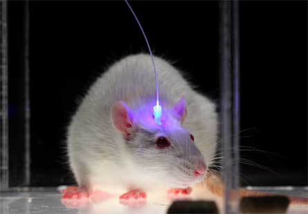
With a flip of a switch, researchers can make a mouse can shed its anxious, shy demeanor. The scientists can dial mouse anxiety up or down by lighting up a very specific connection between two parts of the brain.
The results, reported online March 9 in Nature, “gets us that much closer to understanding how the [anxiety] system works or how it doesn’t work in clinical cases,” says neuroscientist and psychiatrist Kerry Ressler, a Howard Hughes Medical Institute investigator at Emory University in Atlanta who was not involved in the study. The results, he says, will help researchers gain a deeper understanding of circuits in the human brain important for psychiatric disorders.
The new study focused on the amygdalae, a pair of structures buried deep within the brain, one on each side. These bundles of nerve cells are important for emotions, including fear, but it’s been less clear what role this brain region plays in anxiety, which unlike fear doesn’t require a specific trigger.
Researchers led by neuroscientist and psychiatrist Karl Deisseroth, a Howard Hughes Medical Institute investigator at Stanford University, genetically engineered light-sensitive proteins that can turn brain cells on or off in mice, a trick that forms the basis of the growing field of optogenetics (SN: 1/30/10, p. 18). But the researchers added a new twist: Instead of manipulating an entire nerve cell, which would affect all of the cell’s many fingerlike projections that carry information to other cells, the team targeted very specific parts of connections between cells.
A particular connection — the place where cells in the basolateral part of the amygdala connect to the central amygdala — was the anxiety sweet spot, the researchers found. Normally, mice are afraid of wide-open spaces, where they could be nabbed by a cat or a bird. When given a choice in lab tests, mice spend most of the time hunkered down in a platform area with wallsavoiding open areas. But when the connection between these two amygdala neighborhoods was boosted with a burst of light, mice quickly began exploring the formerly frightening areas. When the light was turned off, the mice retreated to the walled areas.
The opposite was also true, the team found. In another experiment, dampening the connection between the two amygdala regions made mice less likely to venture out from the walled areas. “It seems as though within the amygdala, there’s a real-time dial for turning down anxiety,” Deisseroth says.
Having a way to quickly tune anxiety levels is important for animals that have to adjust to new threats, Deisseroth says. “As soon as the organism behaviorally transitions from one environment to another, which can take place in just a couple of seconds, anxiety should be regulated to be reset to the right zone.”
Neuroscientist Elizabeth Bauer of Barnard College in New York City says that other brain regions, some of which are closely connected to the amygdala, probably play a role in tuning anxiety. The specific connection identified in the new study is “definitely an important piece, but of course it’s not the whole story,” she says.
Ressler cautions that extending the results to people might not be straightforward. “We always have to be careful about our translation of how good those models are to what we call human anxiety.” The types of anxiety that he sees in the clinic may be much more complex than what mice experience in experiments, he says.
Tye_Deisseroth_Supplementary Movie from Deisseroth Lab on Vimeo.
Here, a representative mouse from the ChR2:BLA-CeA group is tested on the elevated plus maze during a 15-minute session (played at 10x speed). The session is divided into 3 epochs, and light stimulation is delivered to the BLA terminals in the CeA via an optical fiber only during the second epoch as indicated by the appearance of the blue text detailing light stimulation parameters.


